Zygomatic process of temporal bone
| Zygomatic process of temporal bone | |
|---|---|
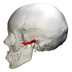 Zygomatic process shown in red. | |
|
Articulation of the mandible. Lateral aspect. (Zygomatic process visible at center.) | |
| Details | |
| Identifiers | |
| Latin | processus zygomaticus ossis temporalis |
| TA | A02.1.06.067 |
| FMA | 52886 |
The zygomatic process of the temporal bone is a long, arched process projecting from the lower part of the squamous portion of the temporal bone. It articulates with the zygomatic bone.
This process is at first directed lateralward, its two surfaces looking upward and downward; it then appears as if twisted inward upon itself, and runs forward, its surfaces now looking medialward and lateralward.
The superior border is long, thin, and sharp, and serves for the attachment of the temporal fascia.
The inferior border, short, thick, and arched, has attached to it some fibers of the masseter.
Surfaces
The lateral surface is convex and subcutaneous.
The medial surface is concave, and affords attachment to the masseter.
Ends
The anterior end is deeply serrated and articulates with the zygomatic bone.
The posterior end is connected to the squama by two roots, the anterior and posterior roots.
- The posterior root, a prolongation of the upper border, is strongly marked; it runs backward above the external auditory meatus, and is continuous with th.
- The anterior root, continuous with the lower border, is short but broad and strong; it is directed medialward and ends in a rounded eminence, the articular tubercle (eminentia articularis).
See also
Additional images
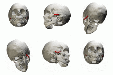 Animation.
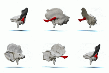 Left temporal bone. Animation.
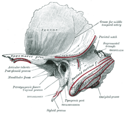 Left temporal bone. Outer surface. Showing the zygomatic process projecting to the left side of the bone.
 Right temporal bone. Inferior surface.
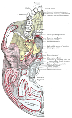 Base of skull. Inferior surface.
- Base of skull. Inferior surface.
References
This article incorporates text in the public domain from the 20th edition of Gray's Anatomy (1918)
External links
| Wikimedia Commons has media related to Zygomatic process of temporal bone. |
- Anatomy diagram: 34257.000-1 at Roche Lexicon - illustrated navigator, Elsevier