Scintillation (physics)
Scintillation is a flash of light produced in a transparent material by the passage of a particle (an electron, an alpha particle, an ion, or a high-energy photon). See scintillator and scintillation counter for practical applications.[1][2]
Overview
The process of scintillation is one of luminescence whereby light of a characteristic spectrum is emitted following the absorption of radiation. The emitted radiation is usually less energetic than that absorbed. Scintillation is an inherent molecular property in conjugated and aromatic organic molecules and arises from their electronic structures. Scintillation also occurs in many inorganic materials, including salts, gases, and liquids.
Scintillation of inorganic crystals
For photons such as gamma rays thallium activated NaI crystals (NaI(Tl)) are often used. For a faster response (but only 5% of the output) CsF crystals can be used.[3]:211
Scintillation of organic scintillators

In organic molecules scintillation is a product of π-orbitals. Organic materials form molecular crystals where the molecules are loosely bound by Van der Waals forces. The ground state of 12C is 1s2 2s2 2p2. In valence bond theory, when carbon forms compounds, one of the 2s electrons is excited into the 2p state resulting in a configuration of 1s2 2s1 2p3. To describe the different valencies of carbon, the four valence electron orbitals, one 2s and three 2p, are considered to be mixed or hybridized in several alternative configurations. For example, in a tetrahedral configuration the s and p3 orbitals combine to produce four hybrid orbitals. In another configuration, known as trigonal configuration, one of the p-orbitals (say pz) remains unchanged and three hybrid orbitals are produced by mixing the s, px and py orbitals. The orbitals that are symmetrical about the bonding axes and plane of the molecule (sp2) are known as σ-electrons and the bonds are called σ-bonds. The pz orbital is called a π-orbital. A π-bond occurs when two π-orbitals interact. This occurs when their nodal planes are coplanar.
In certain organic molecules π-orbitals interact to produce a common nodal plane. These form delocalized π-electrons that can be excited by radiation. The de-excitation of the delocalized π-electrons results in luminescence.
The excited states of π-electron systems can be explained by the perimeter free-electron model (Platt 1949). This model is used for describing polycyclic hydrocarbons consisting of condensed systems of benzenoid rings in which no C atom belongs to more than two rings and every C atom is on the periphery.
The ring can be approximated as a circle with circumference l. The wave-function of the electron orbital must satisfy the condition of a plane rotator:
The corresponding solutions to the Schrödinger wave equation are:
where q is the orbital ring quantum number; the number of nodes of the wave-function. Since the electron can have spin up and spin down and can rotate about the circle in both directions all of the energy levels except the lowest are doubly degenerate.
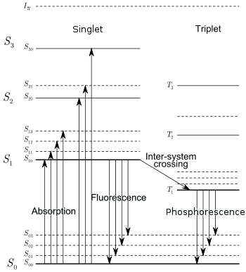
The above shows the π-electronic energy levels of an organic molecule. Absorption of radiation is followed by molecular vibration to the S1 state. This is followed by a de-excitation to the S0 state called fluorescence. The population of triplet states is also possible by other means. The triplet states decay with a much longer decay time than singlet states, which results in what is called the slow component of the decay process (the fluorescence process is called the fast component). Depending on the particular energy loss of a certain particle (dE/dx), the "fast" and "slow" states are occupied in different proportions. The relative intensities in the light output of these states thus differs for different dE/dx. This property of scintillators allows for pulse shape discrimination: it is possible to identify which particle was detected by looking at the pulse shape. Of course, the difference in shape is visible in the trailing side of the pulse, since it is due to the decay of the excited states.