Reflectance difference spectroscopy

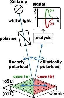
Reflectance difference spectroscopy (RDS) is a spectroscopic technique which measures the difference in reflectance of two beams of light that are shone in normal incident on a surface with different linear polarizations.[2] It is also known as reflectance anisotropy spectroscopy (RAS).[3]
It is calculated as:
and are the reflectance in two different polarizations.
The method was introduced in 1956 for the study optical properties of the cubic semiconductors silicon and germanium.[4] Due to its high surface sensitivity and independence of ultra-high vacuum, its use has been expanded to in situ monitoring of epitaxial growth[5] or the interaction of surfaces with adsorbates.[1] To assign specific features in the signal to their origin in morphology and electronic structure, theoretical modelling by density functional theory is required.
References
- 1 2 May, M. M.; Lewerenz, H.-J.; Hannappel, T. (2014), "Optical in situ Study of InP(100) Surface Chemistry: Dissociative Adsorption of Water and Oxygen", Journal of Physical Chemistry C, 118 (33): 19032, doi:10.1021/jp502955m
- ↑ Peter Y. Yu, Manuel Cardona ,"Fundamentals of Semiconductors"
- ↑ Weightman, P; Martin, D S; Cole, R J; Farrell, T (2005), "Reflection anisotropy spectroscopy", Reports on Progress in Physics, 68 (6): 1251, Bibcode:2005RPPh...68.1251W, doi:10.1088/0034-4885/68/6/R01
- ↑ Aspnes, D. E.; Studna, A. A. (1985), "Anisotropies in the Above-Band-Gap Optical Spectra of Cubic Semiconductors", Physical Review Letters, 54 (17): 1956, doi:10.1103/PhysRevLett.54.1956
- ↑ Richter, W.; Zettler, J.-T. (1996), "Real-time analysis of III--V-semiconductor epitaxial growth", Applied Surface Science, 100--101: 465, doi:10.1016/0169-4332(96)00321-2