LIM domain
| LIM domain | |||||||||
|---|---|---|---|---|---|---|---|---|---|
|
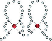
Structure of the 4th LIM domain of Pinch protein. Zinc atoms are shown in grey | |||||||||
| Identifiers | |||||||||
| Symbol | LIM | ||||||||
| Pfam | PF00412 | ||||||||
| InterPro | IPR001781 | ||||||||
| PROSITE | PDOC50178 | ||||||||
| SCOP | 1ctl | ||||||||
| SUPERFAMILY | 1ctl | ||||||||
| CDD | cd08368 | ||||||||
| |||||||||
LIM domains are protein structural domains, composed of two contiguous zinc finger domains, separated by a two-amino acid residue hydrophobic linker.[1] They are named after their initial discovery in the proteins Lin11, Isl-1 & Mec-3.[2] LIM-domain containing proteins have been shown to play roles in cytoskeletal organisation, organ development and oncogenesis. LIM-domains mediate protein–protein interactions that are critical to cellular processes.
LIM domains have highly divergent sequences, apart from certain key residues. The sequence divergence allow a great many different binding sites to be grafted onto the same basic domain. The conserved residues are those involved in zinc binding or the hydrophobic core of the protein. The sequence signature of LIM domains is as follows:
[C]-[X]2–4-[C]-[X]13–19-[W]-[H]-[X]2–4-[C]-[F]-[LVI]-[C]-[X]2–4-[C]-[X]13–20-C-[X]2–4-[C]

LIM domains frequently occur in multiples, as seen in proteins such as TES, LMO4, and can also be attached to other domains in order to confer a binding or targeting function upon them, such as LIM-kinase.
The LIM superclass of genes have been classified into 14 classes: ABLIM, CRP, ENIGMA, EPLIN, LASP, LHX, LMO, LIMK, LMO7, MICAL, PXN, PINCH, TES, and ZYX. Six of these classes (i.e., ABLIM, MICAL, ENIGMA, ZYX, LHX, LM07) originated in the stem lineage of animals, and this expansion is thought to have made a major contribution to the origin of animal multicellularity.[3]
LIM domains are also found in various bacterial lineages where they are typically fused to a metallopeptidase domain. Some versions show fusions to an inactive P-loop NTPase at their N-terminus and a single transmembrane helix. These domain fusions suggest that the prokaryotic LIM domains are likely to regulate protein processing at the cell membrane. The domain architectural syntax is remarkable parallel to those of the prokaryotic versions of the B-box zinc finger and the AN1 zinc finger domains.[4]
References
- ↑ Kadrmas JL, Beckerle MC (2004). "The LIM domain: from the cytoskeleton to the nucleus". Nat. Rev. Mol. Cell Biol. 5 (11): 920–31. doi:10.1038/nrm1499. PMID 15520811.
- ↑ Bach I (2000). "The LIM domain: regulation by association". Mech. Dev. 91 (1–2): 5–17. doi:10.1016/S0925-4773(99)00314-7. PMID 10704826.
- ↑ Koch BJ, Ryan JF, Baxevanis AD (March 2012). "The Diversification of the LIM Superclass at the Base of the Metazoa Increased Subcellular Complexity and Promoted Multicellular Specialization". PLoS ONE. 7 (3): e33261. doi:10.1371/journal.pone.0033261. PMC 3305314
 . PMID 22438907.
 - ↑ Burroughs AM, Iyer LM, Aravind L (July 2011). "Functional diversification of the RING finger and other binuclear treble clef domains in prokaryotes and the early evolution of the ubiquitin system". Mol Biosyst. 7 (1): 2261–77. doi:10.1039/C1MB05061C. PMID 21547297.
